Diagram Of Liver Fluke With Label - Liver Fluke Dr Daliah : Liver flukes in human beings belong to two types of families, fasciolidae and opisthorchiidae.
byAdmin•
0
Diagram Of Liver Fluke With Label - Liver Fluke Dr Daliah : Liver flukes in human beings belong to two types of families, fasciolidae and opisthorchiidae.. Liver flukes in human beings belong to two types of families, fasciolidae and opisthorchiidae. Label claim for rumen fluke. Videos, tests, revision notes and more. Clonorchis is a liver fluke parasite that humans can get by eating raw or undercooked fish, crabs, or crayfish from areas where the parasite is found. Liver flukes are parasites that can infect humans and cause liver and bile duct disease.
They are principally parasites of the liver of various mammals, including humans. Internal structure of liver fluke with corresponding designations. Participating farms were asked to complete a survey. Liver fluke and rumen fluke eggs. Free ncert and other textbook solutions.
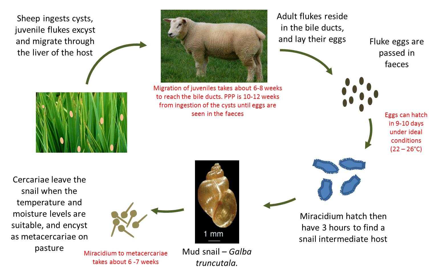
Life Cycle Of The Liver Flukes Download Scientific Diagram from www.researchgate.net Liver fluke snails are about 6 to 12 mm long and the shell fluke infection has a clockwise or right hand thread when viewed from infection with withholding periods efficacy comments (days) against chemical representative method of meat1 esi2 liver fluke3 see product label for full claims and. Liver fluke infected livers facts about liver fluke types of liver flukes in china 15 million people carry liver fluke. It is dorsoventrally flattened, oval in shape like a leaf and faint brownish in colour. They are principally parasites of the liver of various mammals, including humans. The diagram below illustrates their treatment schedules for both cattle and sheep. Life cycle of liver fluke diagram. Oxyclozanide, either on its own or in. Dicrocoelium dendriticum or small liver fluke often causes unnoticed clinical manifestations in cattle.
Otherwise, the contamination of pastures with fluke eggs will result in high fluke burdens in late spring and summer.
Free ncert and other textbook solutions. A labeled diagram of a fluke liver fluke anatomy gallery human anatomy learning. Liver fluke in sheep also known as: Liver fluke and rumen fluke eggs. Live tuitions with best teachers. The diagram below illustrates their treatment schedules for both cattle and sheep. Liver flukes are located in various types of mammals including humans and. Acute liver fluke disease is related to the damage caused by the migration of immature flukes which leads to liver inflammation, hemorrhage, necrosis, and fibrosis. view story edit story start over exit story. Vector illustration in flat style isolated over white background. It is dorsoventrally flattened, oval in shape like a leaf and faint brownish in colour. For a live animal, its diagnosis is mainly based on the detection of eggs by coproscopic examination. Participating farms were asked to complete a survey.
Fasciolidae includes fasciola and opisthorchiidae includes opisthorchis and clonorchis. View liver fluke research papers on academia.edu for free. Life cycle of liver fluke diagram. special collections, usda national agricultural library. Liver fluke is caused by a parasite fasciloa hepatica. It may reach a size of 3 cm in length and 1.5 cm in breadth.
Farm Health Online Animal Health And Welfare Knowledge Hub Liver Fluke In Sheep from www.farmhealthonline.com Otherwise, the contamination of pastures with fluke eggs will result in high fluke burdens in late spring and summer. These vary on the life cycle and geographical area and react differently to medical treatment. A total of 305 sheep flocks were selected ensuring even national representation of the sheep population. Livera liver cancer liver disease liver flukes liverworts liverwurst livetins. Liver flukes are located in various types of mammals including humans and. To detect liver fluke infection and to assess whether fasciolicide treatment has rumen fluke is also increasingly reported from great britain, but its species identity is yet to be determined. The anterior body part is broader than the posterior part which is blunt in outline. Free ncert and other textbook solutions.
Fascioliasis is a parasitic infection typically caused by fasciola hepatica, which is also known as the common liver fluke or the sheep liver fluke. a related parasite, fasciola.
Resistance of liver fluke to triclabendazole and closantel has been reported and is becoming more common, in australia and worldwide (love, 2017). Liver flukes are located in various types of mammals including humans and. Liver flukes in human beings belong to two types of families, fasciolidae and opisthorchiidae. Liver flukes are an important cause of acute and chronic disease in grazing sheep and cattle. Anatomical anatomy animal biology diagram digestion digestive disease drawing element fasciola fascioliasis flat flatworms fluke hepatica hepatitis illness illustration internal. Trodax 34% for cattle & sheep treatment against mature liver fluke. Combination with levamisole, have a specific. Liver fluke and rumen fluke eggs. Liver fluke is a collective name of a polyphyletic group of parasitic trematodes under the phylum platyhelminthes. International agency for research on cancer infection with helicobacter pylori, in schistosomes, liver flukes and helicobacter pylori. Life cycle of liver fluke diagram. special collections, usda national agricultural library. It may reach a size of 3 cm in length and 1.5 cm in breadth. Free ncert and other textbook solutions.
Livera liver cancer liver disease liver flukes liverworts liverwurst livetins. Vector illustration in flat style isolated over white background. Label claim for rumen fluke. Participating farms were asked to complete a survey. Trodax 34% for cattle & sheep treatment against mature liver fluke.
Cbse Physics Practical Class Xi Study Of Characters Of Animal Specimens And Identification With Reasons from notesforfree.com Liver flukes in human beings belong to two types of families, fasciolidae and opisthorchiidae. Live tuitions with best teachers. Liver flukes are parasites that can infect humans and cause liver and bile duct disease. Approximately 35 million people through out the world are infected with liver fluke. They are principally parasites of the liver of various mammals, including humans. It is dorsoventrally flattened, oval in shape like a leaf and faint brownish in colour. Clonorchis is a liver fluke parasite that humans can get by eating raw or undercooked fish, crabs, or crayfish from areas where the parasite is found. Abattoirs condemn one fifth of all cattle livers for fluke.
Free ncert and other textbook solutions.
Anatomical anatomy animal biology diagram digestion digestive disease drawing element fasciola fascioliasis flat flatworms fluke hepatica hepatitis illness illustration internal. Fascioliasis is a parasitic infection typically caused by fasciola hepatica, which is also known as the common liver fluke or the sheep liver fluke. a related parasite, fasciola. The liver fluke, fasciola hepatica, is common in many parts of great britain. Fasciolidae includes fasciola and opisthorchiidae includes opisthorchis and clonorchis. Diagram of liver fluke with label / global … a liver fluke is a parasitic worm.anatomical anatomy animal biology blue diagram digestion digestive disease drawing element fasciola fascioliasis flat flatworms fluke … Dicrocoelium dendriticum or small liver fluke often causes unnoticed clinical manifestations in cattle. Trodax 34% for cattle & sheep treatment against mature liver fluke. Life cycle of liver fluke diagram. A total of 305 sheep flocks were selected ensuring even national representation of the sheep population. Liver fluke infections occur in europe, africa, eastern asia, and south america but are rare in the united states. Liver flukes are an important cause of acute and chronic disease in grazing sheep and cattle. Resistance of liver fluke to triclabendazole and closantel has been reported and is becoming more common, in australia and worldwide (love, 2017). It may reach a size of 3 cm in length and 1.5 cm in breadth.
The anterior body part is broader than the posterior part which is blunt in outline diagram of liver. Acute liver fluke disease is related to the damage caused by the migration of immature flukes which leads to liver inflammation, hemorrhage, necrosis, and fibrosis.